Quantcast
Channel: Press Releases | Heidelberg Engineering Inc.
Viewing all 105 articles
Browse latest View live

International SPECTRALIS Symposium 2017: A Balance of Research Insights and Clinically-Relevant Discussions

$
0
0

Heidelberg, Germany – The 15th International SPECTRALIS Symposium (ISS) took place in Boston, USA, Oct. 13-14, 2017 bringing together SPECTRALIS® enthusiasts from all over the world. Joan W. Miller, MD, Chief of Ophthalmology, Massachusetts Eye and Ear and General Hospital Boston, acted as course director and a faculty of leading experts discussed the latest developments in ophthalmic diagnostic imaging.

Dr. David Brown spoke during the opening session about the effects of microgravity in long-term space flight on the human eye. This subject is a classic at this meeting due to the fact there is a SPECTRALIS on the International Space Station and NASA has relied on this technology for years to monitor the eye-health of astronauts on the ground.

Reflecting on a general trend in ophthalmology, OCT angiography* in a multimodal context of retinal imaging generated significant interest and discussion. Additional highlights of this meeting included sessions on the following:

  • The structure vs. function aspect of AMD (Karl Csaky, MD, PhD, Frank G. Holz, MD)
  • Routine clinical aspects related to the Glaucoma Module Premium Edition (Don Hood, PhD)
  • Innovations in the use of OCT angiography in glaucoma diagnostics (Robert N. Weinreb, MD, Christian Mardin, MD)
  • How the repeatability of the SPECTRALIS platform can create greater clinical efficiencies (Ethan Priel, photographer).

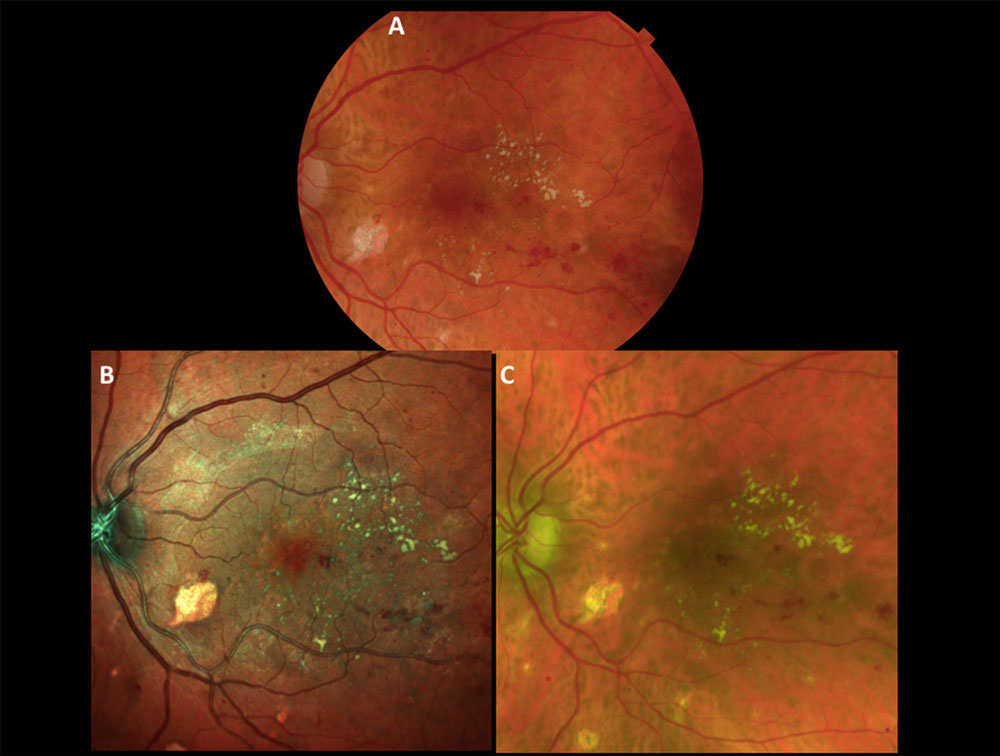
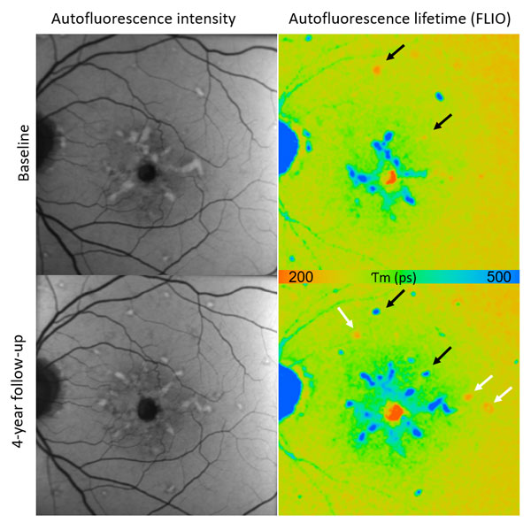
There were presentations about investigational imaging techniques such as: Fluorescence Lifetime Imaging Ophthalmoscopy (FLIO), erythrocyte-mediated angiography or anterior segment outflow imaging. As demonstrated by Dr. Lydia Sauer, of Dr. Paul Bernstein’s research group in Salt Lake City, FLIO might be of particular interest for the diagnosis of Macular Telangiectasia. Dr. SriniVas R. Sadda on his part reported about a study exploring FLIO in the diagnosis of Alzheimer’s disease.

ISS 2017 proved to be a highly interactive meeting with a “scientific marketplace” attendees could visit during breaks as well as frequent Q&A opportunities during the sessions.

The event venue, the State Room, located on the 33rd floor of a high-rise building in downtown Boston, was a highlight by itself. During the breaks ISS attendees could enjoy magnificent views of Boston and its harbor.

At the end of the ISS 2017, it was announced that the 16th International SPECTRALIS Symposium will take place Oct 12-13, 2018 in Bonn, Germany, at the former Plenary Chamber of the German Parliament now known as World Conference Center Bonn.

See ISS videos online

The videos of the ISS 2017 are available on Heidelberg Engineering’s Academy website: https://academy.heidelbergengineering.com/mod/page/view.php?id=824&he_language=en


Dr. Joan Miller ISS 2017
Dr. Joan W. Miller, Chief of Ophthalmology, Massachusetts Eye and Ear and General Hospital Boston acted as course director.
Dr. David Brown Opening Session ISS 2017
Opening session with Dr. David Brown about the effects of microgravity in long-term space flight on the human eye.

*The SPECRALIS OCT Angiography Module is available only in certain countries. Check with your contact person for details.


Heidelberg Engineering Introduces Glaucoma Imaging Atlas

$
0
0

High-Tech OCT Company Shows Commitment to Education with Release of Its Glaucoma Textbook at American Glaucoma Society (AGS) Meeting

Cover of the Glaucoma Imaging Atlas

New York, NYHeidelberg Engineering, a privately-held imaging solutions company focused on advanced diagnostic instruments for eye care professionals, announced today the release of their Glaucoma Imaging Atlas, a comprehensive diagnostic imaging guide for glaucoma assessment and management. The Atlas will serve as the cornerstone of Heidelberg Engineering Academy’s educational programs in glaucoma.

“We are excited to publicly introduce our Glaucoma Imaging Atlas for the first time during American Glaucoma Society’s annual meeting in New York,” said Ali Tafreshi, Director of Clinical Research. “Heidelberg Engineering’s longstanding commitment to high-quality diagnostic imaging has served as a standard in eye care practices and academic institutions across the globe. This project is a culmination of extensive interpretation of OCT images and data by some of the most respected clinicians in the field of glaucoma. The Atlas will be used as a teaching tool to further educate the medical community on the implementation of our OCT technology as an aid in the diagnosis and management of glaucoma, enabling effective, individualized patient care.”

The Glaucoma Imaging Atlas, which features 27 contributors from five countries, includes nearly 30 detailed case studies of patients in various stages of the disease imaged with Heidelberg Engineering’s SPECTRALIS® Glaucoma Module Premium Edition. Heidelberg Engineering will be onsite in New York at AGS’s annual meeting with a copy of the Glaucoma Imaging Atlas available for preview at booth #14.


About SPECTRALIS

The SPECTRALIS is an ophthalmic imaging platform with an upgradable, modular design. This platform allows clinicians to configure each SPECTRALIS to the specific diagnostic workflow in the practice or clinic.

Options include: OCT, multiple scanning laser fundus imaging modalities, widefield and ultra-widefield modules and scanning laser angiography.


About Glaucoma Module Premium Edition

The SPECTRALIS Glaucoma Module Premium Edition combines the proprietary Anatomic Positioning System (APS) with a series of unique scan patterns to assess the optic nerve head, the retinal nerve fiber layer, and the ganglion cell layer. These scan patterns are precisely matched to the characteristics of fine anatomic structures relevant in glaucoma diagnostics.

Dr. Osamah Saeedi receives Heidelberg Engineering 2018 Xtreme Research Award

$
0
0
Dr. Osamah Saeedi receives Heidelberg Engineering 2018 Xtreme Research Award
Osamah J. Saeedi, MD, Associate Professor and Director of Clinical Research at the University of Maryland School of Medicine’s Department of Ophthalmology and Visual Sciences in Baltimore, MD, USA, receives the 2018 Xtreme Research Award from Christopher A. Girkin, MD, MSPH, EyeSight Foundation of Alabama Endowed Chair; Chairman, Department of Ophthalmology, School of Medicine; Chief Medical Officer, Callahan Eye Hospital, University of Alabama in Birmingham, AL.

Heidelberg, Germany – Osamah J. Saeedi, MD, Associate Professor and Director of Clinical Research at the University of Maryland School of Medicine’s Department of Ophthalmology and Visual Sciences in Baltimore, Maryland, USA, received the award for his in vivo imaging of ocular blood flow. Dr. Saeedi presented a lecture entitled “Beyond Angiography: In Vivo Imaging of Ocular Erythrocytes” at the award ceremony, held during ARVO’s Annual Meeting in Hawaii.

The groundbreaking research for which Dr. Saeedi received the Xtreme Research Award is based on a novel technique that allows for labeling of individual erythrocytes (red blood cells) with the fluorescent dye Indocyanine Green (ICG). This technique, combined with dynamic confocal scanning laser imaging, offers the ability to visualize and potentially quantify ocular blood flow in a more granular fashion than ever before. While this research is still in early stages, the dynamics of individual erythrocytes may reveal undiscovered information about the physiological and biological processes at a cellular level within the eye.

“These new discoveries may shed new light on important aspects of ocular hemodynamics such as oxygenation and flow speed, allowing for improvement and/or validation of technologies such as OCT angiography*,” said Ali Tafreshi, Director of Clinical Research at Heidelberg Engineering.

This technique, referred to as erythrocyte-mediated angiography (EMA), is based on the innovative approach of labeling individual red blood cells with the ICG fluorescent dye and then reinjecting them back into the body to enable acquisition of angiograms of the eye. When combined with the video angiography functionality of the SPECTRALIS HRA imaging platform, this novel labeling technique offers the ability to dynamically visualize and monitor the movement and pausing of individual red blood cells in vivo. Dr. Saeedi’s work intends to facilitate a better understanding of the biological processes associated with ocular hemodynamics in a way that incites the identification of new biomarkers for ocular diseases such as glaucoma, AMD and diabetic retinopathy. Although Dr. Saeedi’s main area of interest is glaucoma, his research is laying the groundwork for future studies that may reveal new and important diagnostic parameters for ocular and even systemic diseases.

Other angiography techniques in comparison
Current ocular blood flow imaging techniques are limited in the amount of information that they offer. Conventional dye-based angiography imaging methods are able to visualize ocular blood flow dynamics only in the initial vessel filling phase. Dye-based angiography also mainly offers only 2-dimensional visualization of vasculature. While a new imaging technique, referred to as OCT angiography*, provides non-invasive 3-dimensional visualization of vessel networks, it currently does not offer dynamic information relevant to disease diagnosis. Doppler OCT has been investigated for quantification of ocular blood flow, but this approach has yet to be proven as a reliable and feasible in vivo imaging technique. The novel technique established by Dr. Saeedi may trigger the evolution of studies to evaluate ocular hemodynamics in a more refined fashion than existing methods, addressing some of the current key limitations in this area of research.

Dr. Saeedi’s take
At the award ceremony Dr. Saeedi declared:

“We have developed this novel technique to where it is now: a safe and effective procedure in humans. With better understanding of microvasculature dynamics gained from EMA, we may see the earliest potential changes in conditions such as glaucoma, diabetic retinopathy and age related macular degeneration. This has the exciting potential to open the door to new treatments for these blinding diseases in the earliest stages, a goal we are actively working towards.”

*The SPECTRALIS OCT Angiography Module is available only in certain countries. It has not been cleared by the FDA for use in the United States. Check with your country’s contact person for details on local availability.


Heidelberg Engineering’s SPECTRALIS OCT2 Module Headed to the International Space Station

$
0
0

NASA has been conducting research on the effects of microgravity on vision using the SPECTRALIS imaging platform since 2013.

Franklin, MA. – May 22, 2018 – Heidelberg Engineering, a privately-held imaging solutions company focused on advanced diagnostic instruments for eye care professionals, announced the continuation of the SPECTRALIS space odyssey as the SPECTRALIS imaging platform with the company’s next-generation OCT2 Module is headed for the International Space Station (ISS).

NASA launched the SPECTRALIS with OCT2 Module to the ISS aboard the Antares 230 Cygnus CRS OA-9, also known as Orbital Sciences CRS Flight 9E on May 21, 2018 from Wallops Island, Virginia. The device will be installed on the ISS in late 2018.

“The SPECTRALIS with OCT2 Module uses patented TruTrack Active Eye Tracking,” said Ali Tafreshi, Director of Clinical Research for Heidelberg Engineering. “The technology uses a second laser beam to actively track the eye during OCT scanning to effectively ‘freeze’ the retina and avoid motion artifacts. With this technology, a precise OCT image can be captured, even if the subject blinks or moves.”

NASA’s researchers have used the SPECTRALIS OCT to investigate the effects of a microgravity environment on vision since 2013. While the existing device continues to function normally on the ISS, a next-generation SPECTRALIS with OCT2 Module will optimize acquisition speed and capture more complex scans, while considering use of additional modalities such as OCT angiography*, anterior segment imaging, ultra-widefield fundus imaging and MultiColor.

Heidelberg Engineering’s SPECTRALIS OCT2 Module Headed to the International Space Station
An Orbital ATK Cygnus spacecraft launched from NASA’s Wallops Flight Facility on May 21, 2018.

“Upgrading to the OCT2 Module allows NASA to gain greater understanding of Space Flight Associated Neuro-Ocular Syndrome (SANS), a condition astronauts commonly experience as a result of space flights,” said Alex Huang, MD, PhD, Assistant Professor, Department of Ophthalmology, David Geffen School of Medicine, University of California, Los Angeles. “SANS can cause unilateral and bilateral optic disc edema. In association with the edema, globe flattening, choroidal and retinal folds, refractive error shifts, and nerve fiber layer infarcts have also been noted.”

This research is expanding the exploration of ocular pathology in space and on Earth.

“The SPECTRALIS OCT2 module will allow us to image deeper structures in the eye that are affected by long-term space flight such as the posterior optic nerve head anatomy and the choroidal blood supply to the retina.” said David M. Brown, MD, the retina specialist on the NASA SANS Research & Clinical Advisory Panel and a retinal surgeon at Houston Methodist Hospital in Texas.

“Understanding changes in these structures will be critical for future long-term missions to Mars and have applicability to terrestrial research on both retina disease and glaucoma.”

 
*The SPECTRALIS® OCT Angiography Module is available only in certain countries. It has not been cleared by the FDA for use in the United States. Check with your country’s contact.

Heidelberg Engineering’s SPECTRALIS MultiColor Module Shines at ARVO in Comparisons to Color Fundus Photography

$
0
0

Side-by-side evaluations show MultiColor provides better visualization of certain pathologies

Heidelberg, Germany – Multiple international studies evaluating the sensitivity of SPECTRALIS MultiColor confocal scanning laser ophthalmoscopy versus color fundus photography were presented at the Association for Vision and Research in Ophthalmology (ARVO) 2018 conference in Honolulu, Hawaii. The studies presented at ARVO reported better sensitivity and visualization of a number of pathologies using the SPECTRALIS MultiColor Module in comparison to color fundus photography.

The SPECTRALIS MultiColor Module differs from color fundus photography in that it uses three laser wavelengths simultaneously and a 3.5μm/pixel resolution to provide diagnostic images that show distinct structures at different depths within the retina and clearer images than conventional color fundus photography.

Amit Meshi, UCSD, San Diego, California, USA, et al.1 compared retinal pathology visualization and detection between the SPECTRALIS MultiColor Module, Optos P200 and Topcon TRC-50DX. The authors concluded that while the overall detection rate of retinal pathology was similar across all three devices, a larger area of macular involvement and significantly better epiretinal membrane (ERM) visualization were noted on the SPECTRALIS MultiColor images. A comparison of the three technologies is shown in figure 1.

A study by Ryoh Funatsu, Kagoshima University, Kagoshima-shi, Japan, et al.2 was in agreement with the findings by Amit Meshi et al. and concluded that “evaluation of ERM by MultiColor is more sensitive than color fundus photography” and in addition reported that “MultiColor might be a possible marker for metamorphopsia in ERM patients.”

For the identification of diabetic maculopathy, Obaid Kousha, Ninewells Hospital, London, et al.3 showed that MultiColor combined with SD-OCT provides “effective monitoring” of diabetic maculopathy and that “5 eyes with exudates and severe macular edema requiring urgent intervention were missed by color fundus photography but not by MultiColor.” The benefit of multimodal imaging was highlighted by their finding that “MultiColor, when complemented by SD-OCT, didn’t miss any clinically significant macular edema.”

Hiroto Terasaki, Kagoshima University, Kagoshima-shi, Japan, et al.4 compared fundus images obtained by MultiColor to those obtained using color fundus photography in detecting nerve fiber layer defects. They found that “detection of abnormal findings in the nerve fiber layer by MultiColor was more sensitive than by color fundus photography.”

Find out more about the MultiColor Module for SPECTRALIS at www.spectralis-multicolor.com.

  1. Amit Meshi, UCSD, San Diego, California, USA, et al. Comparison of retinal pathology visualization in multi-spectral scanning laser imaging. Abstract Number: 1501 – C0342, ARVO 2018.
  2. Ryoh Funatsu, Kagoshima University, Kagoshima-shi, Japan, et al. Comparison of color fundoscopy and SPECTRALIS MultiColor on detection of epiretinal membrane and relationship between ERM findings and metamorphopsia. Posterboard Number: C0111, ARVO 2018.
  3. Obaid Kousha, Ninewells Hospital, London, et al. Diabetic maculopathy: Multicolor and SD-OCT versus fundus photography. Posterboard Number: C0111, ARVO 2018.
  4. Hiroto Terasaki, Kagoshima University, Kagoshima-shi, Japan, et al. Diabetic maculopathy: Multicolor and SD-OCT versus fundus photography. Presentation Number: 3444, ARVO 2018


Heidelberg Engineering’s SPECTRALIS MultiColor Module Shines at ARVO in Comparisons to Color Fundus Photography
Figure 1. Images of the same eye taken using A: Topcon TRC-50DX, B: SPECTRALIS MultiColor Module, C: Optos P200. (Amit Meshi, et al. Comparison of retinal pathology visualization in multi-spectral scanning laser imaging. ARVO, 2018).

Heidelberg Engineering prepares leadership team for the future

$
0
0

Heidelberg, Germany – Heidelberg Engineering GmbH announced today changes to the leadership team responsible to consistently deliver the company’s brand promise of excellence and innovation, supported by knowledge and confidence.

Heidelberg Engineering’s new leadership team from left to right in the front row: Managing Directors Arianna and Christoph Schoess, Krysten Williams, Andrea Ritter. Second row from left to right: Ali Tafreshi, Erich Bangert, Claus Gärtner, Dr. Tilman Otto, Kfir Azoulay.

Having worked alongside the founders Dr. Gerhard Zinser and Christoph Schoess for over 23 years, Dr. Tilman Otto will continue to lead R&D. New to the leadership team, Ali Tafreshi who has served in sales, marketing and research capacities in both the US and German organizations for 6 years, will head up Product Management and Clinical Affairs. Dr. Gerhard Zinser carefully handpicked this powerful combination of technical and clinical talent to succeed him. After establishing the UK subsidiary seven years ago and growing into an important force in the market, Krysten Williams will move to the headquarters and support the Marketing and Education efforts. Ali Tafreshi and Krysten Williams will join Dr. Tilman Otto (R&D), Claus Gärtner (Finance, IT and Operations), Kfir Azoulay (Business Development and Healthcare IT), Erich Bangert (Sales and Customer Service) and Andrea Ritter (Human Resources) to form the new leadership team.


“In this leadership team we have so many talented people who have worked with the founders of the company for many years and others who have joined the company because of our ethos of uncompromising integrity,” explained Arianna Schoess, Managing Director of Heidelberg Engineering. “This is the next generation that will carry on the tradition of exploring avenues that combine the company’s core technologies to create innovative, clinically relevant imaging solutions that empower clinicians to improve patient care.”

In this context, the company has parted ways with Managing Director, Dr. Kester Nahen, and thanks him for his important contribution to the success of the company over more than a decade. Arianna and Christoph Schoess will continue to jointly lead the company as Managing Directors in a manner that will support the newly-formed leadership team in Germany to focus on key areas of the business and draw on individual strengths.

As Heidelberg Engineering approaches 30 years of innovation in ophthalmic imaging and ownership passes to the next generation, Arianna Schoess understands the importance of fostering a positive collaborative environment to build a sustainable future for the company.

“Our family business has been built on the mutual trust and respect of a unique partnership between my father and Gerhard, the founders of the company. Their genuine humility has tempered success with self-examination and continuous improvement.” Arianna continues. “This is the legacy that has been left in my care and I want to preserve it. If we uphold these principles, I know that we can continue to be successful.”

Heidelberg Engineering OCT Angiography Module Now Available in the United States

$
0
0

OCTA is a key component of the SPECTRALIS portfolio, delivering on the platform’s upgradeability and multimodal image capabilities.

Franklin, MA – September 17, 2018 – Heidelberg Engineering, a privately-held imaging solutions company focused on advanced diagnostic instruments and IT solutions for eye care professionals, announced today that it has received US Food and Drug Administration (FDA) clearance for its OCT Angiography Module (OCTA). The additional module is now available for new and existing SPECTRALIS® upgradeable diagnostic imaging devices.

“We are excited to announce this FDA clearance as further validation not only of OCTA technology in the industry, but also of our commitment to meeting the highest expectations of our customers,” said Ram Liebenthal, General Manager of Heidelberg Engineering, Inc.

“Our international experience with the SPECTRALIS OCTA Module has confirmed the significance of our live eye tracking and dynamic angiography to acquire clinically relevant data. It also demonstrates our continued dedication to delivering high-resolution images, powerful clinical tools, and a multimodal diagnostic approach to aid eye care professionals in the detection and management of diverse eye conditions, all on a single platform.”

OCTA is a noninvasive imaging technique that provides three-dimensional visualization of perfused ocular vasculature. Combined with structural OCT and dye-based angiography, the OCTA technology is designed to give clinicians a more comprehensive understanding of ocular abnormalities.

“Since the original 510(k) clearance of OCT angiography, there have been significant advancements in the technology and specifically in its clinical applicability, making the validation of the technology a much more rigorous task,” said Ali Tafreshi, Head of Product Management and Clinical Affairs. “For this reason, it was vital to collaborate with respected experts who could clinically validate our OCTA Module to receive 510(k) clearance.”

“The addition of OCT angiography to Heidelberg’s SPECTRALIS multimodal platform allows for the evaluation of retinal and choroidal vascular abnormalities in a noninvasive manner,” said Nadia K. Waheed, MD, MPH, Director of the Boston Image Reading Center and Associate Professor of Ophthalmology, Tufts University School of Medicine, Boston. “I believe OCTA will soon become invaluable in our daily clinical routine for screening and diagnosing chorioretinal disorders.”

“OCTA is an amazing addition to Heidelberg’s already best-in-class OCT retina imaging technology we use in our 12 Houston area clinics to manage diabetic retinopathy, age-related macular degeneration, and other macular diseases,” said David M. Brown, MD.

Dr. Brown is the Director of the Greater Houston Retina Research Center and a retina specialist at Retina Consultants of Houston. Retina Consultants of Houston served as a clinical trial site for this FDA clearance and Dr. Brown served as the principal investigator (PI).

“With the introduction of new technology such as OCTA, we recognize there is a great need for continual learning,” said Mr. Liebenthal. “For this reason, we are fully committed to providing best-in-class training and education through the Heidelberg Engineering Academy to ensure clinicians are empowered to confidently use the technology in daily practice.”


Heidelberg Engineering OCT Angiography Module now available in the United States
Figure 1. Images acquired with the SPECTRALIS OCT Angiography Module of the Superficial Vascular Complex, Deep Vascular Complex, and Avascular Complex of the same patient

Heidelberg Engineering Academy Invites AAO 2018 Attendees to Experience Empowering Educational Sessions in Chicago

$
0
0

Franklin, MA – Heidelberg Engineering, a privately-held imaging solutions company focused on advanced diagnostic instruments and IT solutions for eye care professionals, will host an array of educational activities at the 2018 American Academy of Ophthalmology (AAO) Annual Meeting from October 27th to the 30th in Chicago. The meeting marks the launch of the SPECTRALIS® OCT Angiography (OCTA) Module.

At AAO we are discussing the clinical applications of our recently FDA-cleared OCT Angiography Module. The experience gathered by international groups shows that the combination of our live retinal eye tracking and our distinct OCTA algorithm generates OCTA images of great contrast and resolution that are directly correlated with structural OCT,” said Ram Liebenthal, General Manager of Heidelberg Engineering, Inc. “Additionally, we are pleased to offer a series of short, empowering educational in-booth sessions that feature an impressive lineup of leading clinicians who will offer insight into improving patient care through a multimodal imaging approach.”

These educational sessions will take place in Booth 2236 – South Building, Level 3, Hall A.

In-Booth Educational Sessions on the Advanced OCTA Module for SPECTRALIS

  • Saturday, October 27 at 11:30 am & 3:30 pm
  • Sunday, October 28 at 3:30 pm
  • Monday, October 29 at 11:30 am & 1:30 pm

In-Booth Educational Sessions with Leading Clinicians

Saturday, October 27

  • Tim Steffens, CRA, OCT-C, FOPS; 10:15 am-10:30 am; “OCT2 Technology: How Saving Time Creates Value for Your Practice”
  • Jeffrey G. Gross, MD; 2:15 pm-2:30 pm; “Macular Pucker: A Multi-Colorful Way to See What You Are Missing!”

Sunday, October 28

  • Karl Csaky, MD, PhD; 10:15 am-10:30 am; “The Role of Multimodal Imaging in Real-World Practice”
  • Jeffrey G. Gross, MD; 12:30 pm-12:45 pm; “Macular Pucker: A Multi-Colorful Way to See What You Are Missing!”
  • Sanjay Asrani, MD; 1:15 pm-1:30 pm; “MultiColor Plus OCT: Interface of Glaucoma and Retina”
  • Patrick A. Sibony, MD; 2:00 pm-2:15 pm; “Distinguishing Papilledema from Pseudopapilledema Using OCT”

Monday, October 29

  • Donald C. Hood, PhD; 10:15 am-10:30 am; “Making Optimal Use of SPECTRALIS Glaucoma Module Premium Edition”
  • Alex Huang, MD, PhD; 12:15 pm-12:30 pm; “Imaging to Develop Countermeasures for Visual Deep Spaceflight Challenges and Why it Matters on Earth”

Experience Empowering Educational Sessions in Chicago


SPECTRALIS High Magnification Module Receives FDA Clearance

$
0
0

Similar to a microscope, this module empowers clinicians to see a more magnified and enhanced resolution view of previously imperceivable retinal microstructures

Franklin, MA – Heidelberg Engineering, a privately-held ophthalmic solutions company focused on advanced diagnostic instruments and IT solutions for eye care professionals, announced today it has received US Food and Drug Administration (FDA) clearance for its SPECTRALIS® High Magnification Module. This new module consists of a highly innovative lens and software upgrade that will be available for new and existing SPECTRALIS diagnostic imaging devices.

The SPECTRALIS High Magnification Module elegantly demonstrates the capability of confocal scanning laser ophthalmoscopy (cSLO) to resolve microstructures by reducing intraocular light scattering when imaging ocular structures. The confocal scanning laser ophthalmoscope in the SPECTRALIS platform combines the selectivity of laser light with confocal scanning to provide image detail and clarity not available from fundus photography. Since cSLO imaging minimizes the effects of light scatter, it can also be used effectively even in patients with cataracts. This technology not only offers documentation of clinical findings but also often highlights critical diagnostic details not visible on traditional clinical ophthalmoscopy.

Even when imaging using high-performance cSLO technology, some ocular microstructures may be overlooked in diagnostic images when reviewing scans with larger fields of view. The multi-modality imaging properties of the SPECTRALIS allow the eye care professional to identify a region of interest within the standard field of view and then acquire magnified images at an enhanced resolution to investigate these regions at a microscopic level. The ability to see at a microscopic level enables the eye care professional to customize surgical and treatment regimen for the best possible visual outcome.

“Having a magnified, high-resolution confocal view of ocular structures holds substantial promise,” explains Ali Tafreshi, Head of Product Management and Clinical Affairs for Heidelberg Engineering GmbH.

“The ability to assess ocular microstructures in a magnified view and at an enhanced resolution may provide unique insight into the manner in which diseases progress.”

The SPECTRALIS High Magnification Module will be available for demonstration at the American Academy of Ophthalmology booth 2236 and will be commercially available in December 2018.

~8° High Magnification Module Infrared Confocal Scanning Laser

Regions within SPECTRALIS High Magnification Module images expanded to appreciate the resolution and details of ocular microstructures that can be visualized

SPECTRALIS OCT2 Module Improves Speed and Efficiency on the International Space Station

$
0
0

Research on the effects of microgravity on vision continues with the SPECTRALIS multi-modal imaging platform.

Franklin, MA – Heidelberg Engineering, a privately-held imaging and healthcare IT technologies company focused on ophthalmic diagnostic solutions that empower clinicians to improve patient care, announced that its SPECTRALIS imaging platform with the company’s next-generation OCT2 Module has been successfully installed at the International Space Station (ISS) and is now fully operational.

NASA launched the SPECTRALIS with OCT2 Module to the ISS aboard the Antares 230 Cygnus CRS OA-9, also known as Orbital Sciences CRS Flight 9E, from Wallops Island, Virginia, last May. The new imaging platform was placed into service in late December 2018. The SPECTRALIS with OCT2 Module replaces the SPECTRALIS OCT that has been operational at the ISS since 2013.

“We at Heidelberg are particularly proud of the collaboration with NASA to have our imaging technology be a part of this mission,” said Arianna Schoess Vargas, CEO. “With this advanced OCT technology, it is easier for the astronauts to capture high-quality images in even less time. Working in such an environment, there is significant value placed on efficiency that doesn’t compromise quality.”

SPECTRALIS with OCT2 Module represents the next-generation OCT technology as it provides noticeably faster scan speed, while a more sensitive signal detection enhances image quality and preserves the resolution in the inner retina. Utilizing TruTrack Active Eye Tracking within the OCT2 Module, clinicians are also able to reduce variability between exams. The end results are repeatable and reproducible follow-up exams, higher image quality, improved workflow and a better patient experience with shorter examinations.

“By implementing the next-generation SPECTRALIS OCT2 Module, NASA will be able to take an even deeper dive into understanding Space Flight Associated Neuro-Ocular Syndrome (SANS),” said David M. Brown, MD, the retina specialist on the NASA SANS Research and Clinical Advisory Panel and retinal surgeon at Houston Methodist Hospital in Texas.

“In response to the conditions of long duration space flight, the eyes of almost all of our astronauts experience swelling in the blood supply layers (the choroid) behind the retina and swelling of the optic nerve as it enters the eye. The SPECTRALIS OCT2 Module will enable imaging of these deeper layers of the choroid and optic nerve throughout the space flight and upon the astronauts’ return to Earth’s gravity. Discovering the underlying mechanism of SANS will be mission critical to develop countermeasures necessary for planned spaceflights to Mars and beyond.”

Heidelberg Engineering Introduces the GMPE Hood Glaucoma Report for SPECTRALIS OCT

$
0
0

New feature of the Glaucoma Module Premium Edition offers an intuitive and accurate approach to aid diagnosis and management of glaucoma

Franklin, USA – Heidelberg Engineering, a privately-held imaging and healthcare IT technologies company focused on ophthalmic diagnostic solutions that empower clinicians to improve patient care, announced today that the Hood Glaucoma Report is now available with a software update within the SPECTRALIS OCT Glaucoma Module Premium Edition (GMPE).

The GMPE Hood Glaucoma Report highlights essential diagnostic information in an intuitive layout that enables a quick, yet comprehensive assessment. Based on the diagnostic approach developed by Donald C. Hood, PhD, this report also accentuates the importance of high-resolution OCT B-scans and the unique anatomy of each eye, in the routine clinical diagnostic regimen.

“With the NSTIN plot, the temporal quadrant of the retinal nerve fiber layer is displayed in the center of the B-scan. This portion of the disc is crucial for everyday visual function, and identification of abnormal thinning in this region is essential for managing glaucoma.” said Dr. Hood.

Furthermore, this report allows clinicians to visualize functional and structural measurements along with high-resolution OCT B-scans and relate this information to 10-2 and 24-2 visual field points. Taking advantage of the GMPE Anatomic Positioning System (APS), which tailors scan placement and orientation to each patient’s individual anatomy, the report is optimized to serve as an intuitive and robust diagnostic aid. The unique semi-automated APS technology increases the precision and accuracy of results by ensuring that all glaucoma scans are anatomically aligned with the reference database and account for the individual configuration of axons in each eye.

“The GMPE Hood Glaucoma Report makes full use of the high-quality images and precise diagnostic information provided by the SPECTRALIS to make it even more convenient for clinicians to identify subtle changes and make confident clinical decisions,” stated Ali Tafreshi, Head of Product Management and Clinical Affairs.

GMPE Hood Glaucoma Report
Figure 1. The GMPE Hood Glaucoma Report

Heidelberg Engineering’s SPECTRALIS Technology Spotlighted in Multiple Presentations at ARVO 2019

$
0
0

Participants are invited to attend the Company’s annual Xtreme Research Award Lecture

Vancouver, B.C. – April 26, 2019 – Heidelberg Engineering, a privately-held company providing diagnostic solutions that empower eye care professionals to improve patient care, proudly announced that its multimodal imaging platform, SPECTRALIS, will be strongly represented at the 2019 Association for Research in Vision and Ophthalmology (ARVO) conference, to be held April 28 – May 2 in Vancouver, British Columbia. The SPECTRALIS imaging platform, which offers high-resolution visualization of ocular structures and vasculature, plays an integral role in multiple clinical research studies being presented at ARVO.

Heidelberg Engineering encourages meeting attendees to experience the innovative applications of SPECTRALIS by attending relevant poster and paper presentations (partial list included below). Additionally, Heidelberg Engineering’s 2019 Xtreme Research Award lecture will be held at 7:30 PM Monday, April 29 at Pan Pacific Vancouver, 300-999 Canada Place. The recipients of this year’s award, Lydia Sauer, MD, and Chantal Dysli, MD, PhD, will present their compelling fluorescence lifetime imaging ophthalmoscopy (FLIO) research. To learn more and register, please visit: https://www.heidelbergengineering.com/us/arvo-xtreme/.

“Through our continual technological advancements and longstanding collaborations with innovators in the ophthalmic field, Heidelberg Engineering has exhibited its ongoing commitment to meaningful novelties and quality images. Accordingly, we are proud to play a large role in research being presented at ARVO 2019,” said Mr. Ali Tafreshi, Head of Product Management and Clinical Affairs. “We invite the ophthalmic community to learn more about our latest technology for both the anterior and posterior segments, on-site in Vancouver”.

Recent Innovations to Be Presented at Booth 1117

SPECTRALIS OCT Angiography Module
The OCTA Module for SPECTRALIS delivers high-resolution, non-invasive imaging of ocular vasculature. Combined with the precision of TruTrack Active Eye Tracking, the OCTA Module enables visualization of fine capillary networks in great detail.

SPECTRALIS High Magnification Module (HMM)
This new module, which consists of an innovative lens and software, elegantly demonstrates the capability of confocal scanning laser ophthalmoscopy (cSLO) to resolve ocular microstructures by reducing intraocular light scattering. The detail seen in HMM images may provide novel insights into the pathogenesis and progression of ocular diseases.

ANTERION®*
Heidelberg Engineering has optimized the longer wavelength and lower sensitivity roll-off of swept-source OCT technology for imaging the anterior segment. ANTERION offers topography, tomography, anterior segment metrics and axial length in one upgradeable, modular device. The stunning swept-source OCT images empower clinicians with visual confirmation of the reliability and accuracy of the measurements.

*ANTERION is not cleared for sale in Canada or the United States.

Paper Presentations Featuring SPECTRALIS*

Wednesday, May 1, 2019

  • 11:15 AM ARVO Ballroom: Optic nerve head (ONH) hypercompliance and blood flow autoregulation dysfunction detected by OCT-angiography (OCTA) in early-state experimental glaucoma (EG)
  • 11:30 AM East 2/3: Reproducibility of retinal erythrocyte velocity using human erythrocyte-mediated angiograms
  • 10:30 AM ARVO Ballroom: Optic nerve head morphological changes over a 12-hour period in a seated vs. head-down tilt posture

Poster Presentations Featuring SPECTRALIS*

Sunday, April 28, 2019

8:00 AM – 9:45 AM

  • 189 – A0598: Optimization and validation of a novel technique for in vivo imaging of FITC-dextran labelled erythrocytes in non-human primates

1:00 PM – 2:45 PM

  • 662 – B0140: Extracellular Matrix Tissue content of the neuroretinal rim differs in healthy and glaucomatous eyes

Monday, April 29, 2019

8:15 AM – 10:00 AM

  • 1496 – A0180: Automatic segmentation of retinal capillaries in adaptive optics perfusion images using a convolutional neural network

Tuesday, April 30, 2019

8:45 AM – 10:30 AM

  • 2976 – A0259: Monocyte-related biomarkers of intermediate age-related macular degeneration
  • 3091 – A0434: Factors Affecting the variability of vessel width measurements within individual vessels in OCT angiography
  • 3087 – A0430: Variability and repeatability of retinal vessel caliber measurements with optical coherence tomography angiography

11:45 AM – 1:30 PM

  • 3619 – B0033: Correlation of novel macular structural and vascular abnormalities in familial exudative vitreoretinopathy

Wednesday, May 1, 2019

3:00 PM – 4:45 PM

  • 5596 – B0487: Optic nerve head prelaminar tissue schisis: a sign of glaucomatous deformation?

Thursday, May 2, 2019

8:00 AM – 9:45 AM

  • 5738 – A0133: Determination of absolute diastolic erythrocyte velocities in peripapillary arterioles and venuoles of human subjects using erythrocyte mediated angiography

Please visit us during ARVO at booth #1117.

*This is a partial list of abstracts that include SPECTRALIS.

Dr. Chantal Dysli and Dr. Lydia Sauer receive Heidelberg Engineering Xtreme Research Award 2019

$
0
0
Dr. Chantal Dysli (left) and Dr. Lydia Sauer (right) at the Xtreme Research Award ceremony.

Heidelberg, Germany – Dr. Chantal Dysli (Inselspital University Hospital Bern, Switzerland) and Dr. Lydia Sauer (Moran Eye Center of the University of Utah, USA) are the joint winners of this year’s Xtreme Research Award. The two ophthalmologists have earned this recognition due to their impressive Fluorescence Lifetime Imaging Ophthalmoscopy (FLIO) research and impactful peer-reviewed manuscripts. The award ceremony and lecture were held at ARVO’s Annual Meeting in Vancouver (Canada).

FLIO1 is a novel, non-invasive imaging modality able to detect early metabolic change preceding morphological changes in the retina. It provides in-vivo autofluorescence lifetime measurements of natural retinal fluorophores upon laser excitation.

Dr. Dysli and Dr. Sauer have been involved in numerous studies demonstrating that many retinal pathologies display distinct metabolic patterns in FLIO images before any other diagnostic imaging method can identify structural signs of disease. FLIO detects changes related to AMD, albinism, Alzheimer’s disease, diabetic retinopathy, macular telangiectasia type 2, retinitis pigmentosa, and Stargardt’s disease . Some of these changes can be detected in healthy eyes, serving as potential early indicators of retinal disease. Other changes in already affected eyes seem to reveal disease progression. Once the technology becomes widely available, the diagnostic information provided by FLIO images could be used for early diagnosis and therapy monitoring.

FLIO in Stargardt’s disease: Representative autofluorescence intensity and lifetime image at baseline and at 4 years follow up (long spectral channel 560-720nm). Black arrows highlight flecks with short autofluorescence lifetimes (red) at baselines and a change to long fluorescence lifetimes (blue) at follow up. White arrows indicate new flecks with short lifetimes at follow up. Images courtesy of Dr. Chantal Dysli.

Dr. Dysli and Dr. Sauer have been a driving force in the clinical development and validation of fluorescence lifetime imaging as a diagnostic aid in the detection and management of various ocular and systemic conditions. Their clinical research paves the way for functional ocular imaging as the means to advance the field towards earlier detection of diseases.” said Ali Tafreshi, Head of Product Management and Clinical Affairs at Heidelberg Engineering.

At the award ceremony, Dr. Dysli stated: “FLIO is a fascinating novel technique opening a new dimension in retinal imaging by providing information about basic pathophysiological mechanisms within the retina. For me, it has been very inspiring to follow the development of FLIO from bench to bedside and to see the progress and future clinical applications of such an innovation”.

Dr. Sauer said: “FLIO is an exciting technology to work with. In-vivo metabolic imaging could become a new clinical milestone in our field, so we are delighted to be part of its development. The FLIO technology is useful in the diagnosis of many retinal diseases and as our research attests, it detects changes in the eye even before damage is visible with conventional imaging techniques. FLIO’s ability to show metabolic change before actual damage manifests could also lead to earlier interventions. I feel honored to be a part of this exciting research.

Heidelberg Engineering strengthens its R&D and technology leadership team

$
0
0
Dr. Tilman Otto (left) has taken on the role of Head of Technology Management for Ophthalmic Devices and Dr. Christian Odaker (right) has joined as Head of Research and Development.

Heidelberg, Germany – Heidelberg Engineering is strengthening its R&D and technology management to continue to deliver clinically-relevant diagnostic solutions and breakthrough ophthalmic imaging technologies. Dr. Tilman Otto has been appointed Head of Technology Management for Ophthalmic Devices and Dr. Christian Odaker joins the company as Head of Research and Development.

Heidelberg Engineering is responding to the increasing demands of technology development and the growth and diversification in its own R&D team with this high-caliber management setup. The new organizational structure as well as the company’s heavy investment in R&D over the past five years, are geared towards maintaining the position as provider of innovative ocular imaging technologies that stand the test of time and set new standards for patient care.

With immediate effect, Dr. Tilman Otto, who has been with Heidelberg Engineering for over 20 years, is taking on the role of Head of Technology Management for Ophthalmic Devices. He will focus on planning and optimizing disruptive innovation and delivering cutting-edge technology to further advance the company’s position as a technological leader.

Dr. Christian Odaker has joined the company as Head of Research and Development, with a strong background in informatics and experience leading a multi-disciplinary team at a successful and fast-growing software company in the field of virtual prototyping.

Having worked side by side with our late co-founder Dr. Gerhard Zinser for over 20 years and playing a major role in the development of our patented TruTrack Active Eye Tracking technology, Tilman’s passion is for innovative applications of technology. In his new role, he will concentrate on rapidly evolving technologies and continue to revolutionize ophthalmic diagnostics in close collaboration with leading scientists,” said Arianna Schoess Vargas, Managing Director at Heidelberg Engineering.

On the other hand, Christian’s passion for leadership, his respect for the technical expertise of our team, as well as his proven track record at delivering optimal results make him the right person to drive our R&D initiatives. Together, Tilman and Christian will prioritize and focus R&D efforts on technology opportunities to sustain our industry leadership and future success.

Heidelberg Engineering appoints Christopher Mody as Clinical Director

$
0
0
Chris Mody at ISS
Christopher Mody, Clinical Director of Heidelberg Engineering at the recent International SPECTRALIS Symposium in Seoul, South Korea.

Heidelberg, Germany – In this new role, Christopher Mody, an ophthalmic scientist with 30 years clinical ophthalmology experience in the public sector and 7 years at Heidelberg Engineering UK, will be facilitating the exchange of scientific information between the global ophthalmic community and the company.

From its inception almost 30 years ago, Heidelberg Engineering has worked closely with scientists and clinicians to develop clinically relevant innovative solutions. By appointing Christopher Mody as Clinical Director, the company further strengthens its close ties with the medical community with the goal of identifying and translating clinical needs into the company’s Product Management and Research and Development functions.

“Chris Mody is ideally suited to support our translational science program designed to deliver comprehensive, clinically relevant solutions to our customers. His knowledge will continue to strengthen our research alliances, while his passion for education will enhance the company’s role in advancing patient care.”, said Ali Tafreshi, Head of Product Management and Clinical Affairs at Heidelberg Engineering.

“When I received my first HRT and one of the very first HRAs in Sheffield many years ago, I immediately realized that they were essential to improving our service. With their accuracy and repeatability, we could make a difference for patients. When I joined the Heidelberg Engineering UK team, I witnessed daily how the dynamic diagnostic information that our customers obtain empowers them to deliver better care,” said Christopher Mody, Clinical Director at Heidelberg Engineering. “In my new global role, I will devote myself to the exchange of accurate intelligence between the ophthalmic community and our company for the development of innovative solutions that continue to fulfill clinical needs,” he concludes.

The list of Christopher Mody’s career achievements is long. Starting off as a graduate in Ophthalmic Science and Technology, he went on to become a Clinical Service Manager at the Ophthalmic Physiological Sciences Service of Sheffield Teaching Hospital, where he worked for 24 years. Responsible for one of the busiest ophthalmic departments in the UK, he successfully implemented a healthcare IT infrastructure to deliver paperless working and virtual clinics. Parallel to that, he acted as a consultant to The Royal College of Ophthalmologists and the Department of Health for the design of national occupational standards and for the development of ophthalmic and vision science university curriculum for the National Health Service. As part of the Heidelberg Engineering UK team, he became one of the leading figures of the Heidelberg Engineering Academy raising the standard of imaging education and facilitating numerous significant research projects.

In his new position, Christopher Mody will influence the clinical direction of the entire Heidelberg Engineering product portfolio and the integration of third-party solutions. An example of the tasks ahead of him is the further development and validation of an enhanced diagnostic toolbox for early detection and management of glaucoma; employing state-of-the-art imaging technologies and traditional parameters to deliver multifaceted clinical evaluation of the disease.


New high-caliber publication validates macular pigment assessment with SPECTRALIS

$
0
0
Dr. Marina Green-Gomez and Prof. John M. Nolan during a patient examination
Dr. Marina Green-Gomez and Prof. John M. Nolan, from the Nutrition Research Centre Ireland, School of Health Science, Waterford Institute of Technology in Ireland during a patient examination with the investigational SPECTRALIS Macular Pigment Optical Volume* (MPOV).

Heidelberg, Germany – The paper “Standardizing the Assessment of Macular Pigment Using a Dual-Wavelength Autofluorescence Technique”, published in Translational Vision Science & Technology in December 2019, demonstrates how the investigational SPECTRALIS® Macular Pigment Optical Volume* (MPOV) measurement provides a comprehensive and objective evaluation of the macular pigment (MP) profile.

Macular pigment has been confirmed as an important dietary variable to enhance and protect visual function, with implications for patients with AMD and other retinal pathologies1. A challenge for this research has been to standardize the assessment of macular pigment. Previous techniques have been limited because of their inability to provide a full image, their subjectivity and high variability.

This new research work validates the assessment of macular pigment with the investigational dual-wavelength autofluorescence technique of the SPECTRALIS MPOV and introduces a benchmark for the clinical setting. The authors, Marina Green-Gomez, Paul S. Bernstein, Christine A. Curcio, Rachel Moran, Warren Roche and John M. Nolan, state that the SPECTRALIS MPOV “should be considered a preferred metric for the assessment of macular pigment”2.

The corresponding author, Prof. John M. Nolan, from the Nutrition Research Centre Ireland, School of Health Science, Waterford Institute of Technology in Ireland said:

“Now that the importance of macular pigment has been verified by substantial clinical studies with positive implications for enhancing visual performance for all populations, including patients with age-related macular degeneration; accurate, validated and standardized measurement of human macular pigment now represents a unique opportunity for eye care. The measurement of macular pigment using the Heidelberg Engineering SPECTRALIS represents the new gold standard for objective assessment of this pigment, and I am sure will empower clinicians to do more for their patients as we prepare for a new standard of vision beyond 2020!”

Beyond proposing that the dual-wavelength autofluorescence technique of the SPECTRALIS MPOV becomes the gold standard for macular pigment assessment, the authors envision the standardization of macular pigment assessment across research studies and clinical settings as the foundation for normative databases to be created.

For further details, see this direct link to the publication2

Read Publication

*SPECTRALIS MPOV is currently under development and not available for sale.

1 Bernstein PS, Li B, Vachali PP, et al. Lutein, zeaxanthin, and meso-zeaxanthin: the basic and clinical science underlying carotenoid-based nutritional interventions against ocular disease. Prog Retin Eye Res. 2016;50:34–66.

2 Green-Gomez M, Bernstein PS, Curcio CA, Moran R, Roche W, Nolan JM. Standardizing the assessment of macular pigment using a dual-wavelength autofluorescence technique. Translational Vision Science & Technology December 2019, Vol.8, 41. doi: https://doi.org/10.1167/tvst.8.6.41

Heidelberg Engineering brings back in vivo corneal microscope due to high demand of ophthalmology market

$
0
0

Heidelberg, Germany – Heidelberg Engineering, the industry leader in ophthalmic diagnostic imaging, announces the reintroduction of its HRT3 Rostock Cornea Module (RCM) globally to offer in vivo corneal confocal microscopy in its anterior segment product portfolio, supporting cornea specialists worldwide to further improve patient care.

HRT3 RCM is a compact ophthalmic device that uses confocal scanning laser microscopy to provide high-resolution images of the cornea, the conjunctiva or the limbus at the cellular level. It capitalizes on the proven technology that has differentiated Heidelberg Engineering as an innovator in both the posterior and the anterior segment market for 30 years. The company collaborated with the research group of Prof. Dr. Rudolf Guthoff at the University of Rostock, Germany, to develop a dedicated lens and software application for the HRT platform that enables in vivo microscopy of the cornea.

The resulting HRT3 RCM provides unique en face images of corneal cells and structures. It scans the cornea with a field of view of up to 400 x 400 μm, resulting in high-resolution images that allow the user to navigate through all corneal layers – including the subdifferentiation of various epithelial cell layers –, identify keratocytes subpopulations and even visualize the corneal subbasal nerve plexus. This comprehensive in vivo assessment of the cornea can aid clinicians in the diagnosis and monitoring of corneal pathologies, pre- and post-surgery evaluation, the assessment of dry eye disease, and in the evaluation of corneal nerve structure in diabetic patients.
Its unprecedented imaging capability has made the HRT3 RCM an ideal device for scientific research, which has resulted in numerous publications1. HRT3 RCM has also proven its relevance as a diagnostic platform for daily clinical practice. With growing demand from the clinical evidence base and global market, Heidelberg Engineering decided to reintroduce the in vivo microscope as part of its commercial portfolio.

“The relaunch of the HRT3 RCM is exciting as this unique imaging device aids the ophthalmologist in the diagnosis of corneal diseases by allowing imaging at a cellular level. The high-resolution images have been pivotal for visualization of corneal infections such as acanthamoeba and fungi, as well as quantification of corneal nerves in patients with diabetes mellitus and dry eye disease. The HRT3 RCM is a very promising tool for both the clinician wanting to make a diagnosis in the clinic, as well as researchers interested in the cellular basis of disease, thus facilitating in vivo histology”, says Dr. Jaya Chidambaram, BSc (Hons), MBBS, MRCOphth, PhD, Senior Lecturer, University of Manchester, United Kingdom.

HRT3 RCM will be relaunched in Spring 2020, including a new headrest for corneal assessment. It will also integrate into the company’s HEYEX 2 image management software environment, which enables new functionalities for streamlined workflows, such as automated report generation and archiving, or drag-and-drop exports for paperless data sharing.

“In addition to the contribution the HRT3 RCM has made to scientific research, there is an unmet clinical need for high-resolution in vivo corneal microscopy. We have decided to renew the HRT3 RCM globally both to advance science and improve patient care,” explains Erich Bangert, Vice President of Sales & Service, Heidelberg Engineering.

With its ability to investigate the cornea at the cellular level, HRT3 RCM is expected to empower clinicians in fast-growing clinical areas such as dry eye disease, infectious corneal diseases, corneal dystrophies, diabetic neuropathy, as well as refractive and corneal treatment surgery.

Kindly provided by Prof. R. Guthoff, MD, Prof. J. Stave, PhD, A. Zhivov, MD, Rostock/Germany

1  Villani E, Baudouin C, Efron N, Hamrah P, Kojima T, Patel S V, Pflugfelder S C, Zhivov A, Dogru M. “In Vivo Confocal Microscopy of the Ocular Surface: From Bench to Bedside”. Current Eye Research. 2014; 39(3). Link: https://www.ncbi.nlm.nih.gov/pmc/articles/PMC3960287/

Prof. John M. Nolan receives Heidelberg Engineering Xtreme Research Award 2020

$
0
0

Heidelberg, Germany – Prof. John M. Nolan, from the Nutrition Research Centre Ireland, School of Health Science, Waterford Institute of Technology (Ireland), is the recipient of the Xtreme Research Award 2020 for his research related to macular pigment for vision health and function, as well as for his recent work on how to use macular pigment as a biomarker for brain nutrition and its connection to cognitive function and Alzheimer’s disease.

Prof. Nolan has an impressive research track record focusing on the role of macular pigment (lutein, zeaxanthin and meso-zeaxanthin) for visual and cognitive function. With the Xtreme Research Award 2020, Heidelberg Engineering recognizes his efforts to verify the importance of macular pigment for visual performance in both healthy individuals and AMD patients, standardizing these measurements with an investigational SPECTRALIS Macular Pigment Optical Volume* Module. It also acknowledges his drive to link macular carotenoids to cognitive function and brain health, with the goal of identifying ways to reduce the risk of Alzheimer’s disease.

Prof. Nolan said: “It is a tremendous honor for me to accept this award from Heidelberg Engineering, which is a result of over two-decades of work studying this nutritional pigment in the eye. I am most grateful to my team for their continued commitment to our research questions, and this award is a true reflection of their achievements. Science is only as good as its measurements, and thankfully we now have access to gold standard technology such as the Heidelberg Engineering SPECTRALIS.

The future of our research and its successful translation and benefits to eyecare will be driven by this innovative technology. My vision is that routine measurement of macular pigment in the clinic setting will be part of best practice and uniquely offered by the SPECTRALIS.”

Until recently, it was difficult to standardize the assessment of macular pigment since previous techniques were unable to provide a full image and measurement results were both subjective and highly variable. Prof. Nolan is one of the leading researchers that have contributed to the establishment of a comprehensive and objective evaluation of the macular pigment profile for the benefit of patients worldwide1.

Invitation to an Xtreme Research Award Webinar

Due to this year’s ARVO cancellation, the traditional Xtreme Research Award lecture will take place online as an Xtreme Research Award Webinar with Prof. Nolan: “Macular Pigment Assessment with SPECTRALIS”, Wednesday, May 13, 2020 3:00 PM – 4:00 PM
(CEST).
Heidelberg Engineering invites you to join by registering under: https://www.heidelbergengineering.com/us/events/xtreme-research-award-webinar-11503311/

*SPECTRALIS MPOV is currently under development and not available for sale.

1 Green-Gomez M, Bernstein PS, Curcio CA, Moran R, Roche W, Nolan JM. Standardizing the assessment of macular pigment using a dual-wavelength autofluorescence technique. Translational Vision Science & Technology December 2019, Vol.8, 41. doi: https://doi.org/10.1167/tvst.8.6.41https://doi.org/10.1167/tvst.8.6.41

New Scientific Director strengthens Heidelberg Engineering product development

$
0
0
Charles Reimann
Charles Reisman, newly appointed Scientific Director at Heidelberg Engineering.

Heidelberg, Germany – Charles Reisman has joined Heidelberg Engineering’s global management team as Scientific Director. In this newly created role, he will be responsible for the clinical research plan that directly feeds into the product development pipeline.

Heidelberg Engineering is strengthening its Clinical Research team with the addition of Charles Reisman as Scientific Director. Charles is an accomplished clinical validation expert with decades of ophthalmic industry and other high-technology expertise. He holds an Electrical Engineering degree from the Massachusetts Institute of Technology (MIT) and several patents on signal and image processing methodologies within the field of ophthalmic imaging. He has published in numerous distinguished peer-reviewed scientific journals and book chapters and is also well versed in deep learning and artificial intelligence.

In his new position, Charles will focus on the strategic leadership of Heidelberg Engineering’s clinical research and trial efforts, ultimately geared towards executing the company’s product development pipeline. For that purpose, he will not only work closely with the Research and Development, Clinical and Regulatory Affairs teams but primarily with external researchers from across the world.

“I look forward to contributing to Heidelberg Engineering’s product development goals within our heavily regulated environment. I am particularly inspired by the collaborations with leading clinicians and researchers across the world as well as the impressive team of R&D experts at Heidelberg Engineering, that are among the brightest in the industry,” said Charles Reisman, newly appointed Scientific Director.

“Charles brings excellent credentials and a deep understanding of the ophthalmic research and development landscape. We are confident that his expertise will help us bring more and even better products to the market. We are specifically focused on early detection of diseases by supporting the confident identification of clinical biomarkers,” said Arianna Schoess Vargas, Managing Director of Heidelberg Engineering.

From its inception in 1990, Heidelberg Engineering has collaborated with scientists, clinicians and industry to develop innovative products that deliver clinically relevant benefits. With the appointment of Charles Reisman as Scientific Director, the company signals a renewed commitment to this guiding principle.

The post New Scientific Director strengthens Heidelberg Engineering product development first appeared on Corporate US.

Heidelberg Engineering Emphasizes Anterior Segment Focus under New Clinical Director

$
0
0
Steve Thomson, newly appointed Clinical Director for the Anterior Segment at Heidelberg Engineering

Heidelberg, Germany – Steve Thomson joins Heidelberg Engineering as Clinical Director to further strengthen the Anterior Segment product development team. He will concentrate on the integration of data and images into increasingly efficient and effective anterior segment workflows for clinical practice.

Steve Thomson assumes his new role as Clinical Director – Anterior Segment effective January 1, 2021. He has a background in ophthalmic imaging followed by more than 30 years’ professional experience in the ophthalmic industry. Steve is a knowledgeable and respected anterior segment expert with an in-depth comprehension of clinical ophthalmology, as well as all the diagnostic and surgical technologies involved.

Steve’s focus will be on the dynamic visualization of anterior segment diagnostic information, both in terms of imaging and data, to empower clinicians, scientists and administrators, as well as inform AI solutions to improve patient outcomes. For that purpose, he will work in particularly close cooperation with the new Scientific Director, Charles Reisman, as well as the newly promoted Medical Scientific Liaison – Anterior Segment, Richard Cornwell who now takes on a global role.
“Heidelberg Engineering has set extremely high standards for ophthalmic imaging to the benefit of so many patients across the world. I am excited to be part of this talented team and contribute to the development of ANTERION and other complementary anterior segment solutions with the potential to enhance the biometric and imaging data that is so vitally important to clinicians and surgeons” said Steve Thomson, newly appointed Clinical Director – Anterior Segment at Heidelberg Engineering.

“As a company, we have ambitious plans to shape the anterior segment aspect of clinical routines in a similar way to what we have achieved in the posterior segment. Thanks to his anterior segment expertise, strong relationships with clinicians and researchers and decades of experience, we are convinced that Steve is ideally placed to demonstrate how high-quality imaging combined with comprehensive data can foster confident treatment decisions and transform clinical workflows,” said Arianna Schoess Vargas, Managing Director of Heidelberg Engineering.

In cooperation with the entire product development team, Steve Thomson will pursue Heidelberg Engineering’s ethos of collaboration with scientists, clinicians and industry to develop innovative products that deliver clinically relevant benefits.

Note: ANTERION is not available for sale in the US.

The post Heidelberg Engineering Emphasizes Anterior Segment Focus under New Clinical Director first appeared on Corporate US.
Viewing all 105 articles
Browse latest View live